Anatomy Of Ribs And Chest - Bones in the body Skeletal system 206 bones in the body ... - The second most common chest wall abnormalities that we see on a cxr are metastases in vertebral bodies and ribs.
Anatomy Of Ribs And Chest - Bones in the body Skeletal system 206 bones in the body ... - The second most common chest wall abnormalities that we see on a cxr are metastases in vertebral bodies and ribs.. Spiral ct of thoracic inlet. True, false and floating ribs are denoted. And as you might guess from the word major, it makes up the majority of the chest muscle mass. Powerful muscles that move the head and arms attach to these bones as well. The second most common chest wall abnormalities that we see on a cxr are metastases in vertebral bodies and ribs.
Ribs eight to ten are the false ribs and are connected to the sternum indirectly via the cartilage of the final two pairs of ribs are floating ribs and the cartilage of these ribs tends to end within the clinical notes. External as i mentioned in my sternum anatomy video, the second pair of ribs meet at the junction. Identify the following structures on the lateral chest radiograph: Chest blunt trauma (cbt) and the resultant rib fractures often lead to thoracic collapse. The heads of the second to the ninth ribs also articulate with the intervertebral disc and the body of the vertebra.
Anatomy Of The Ribs And Sternum | MedicineBTG.com from medicinebtg.com Twelve pairs of ribs extend laterally and anteriorly from the thoracic vertebrae to meet at or near the sternum. The second most common chest wall abnormalities that we see on a cxr are metastases in vertebral bodies and ribs. The rib cage also anchors the bones of the head, neck, shoulders, and arms to the trunk of the body. Terms in this set (53). The chest anatomy includes the pectoralis major, pectoralis minor and the serratus anterior. The first seven ribs attach to the sternum directly and are called true ribs. ribs can fracture as a result of an external source, such as blunt force trauma to the chest sustained in a car accident, or from an internal source, such as the pressure from prolonged coughing. Anatomy of the chest and the lungs: Attach directly to sternum.false ribs:
The purpose of this study was to explore the effect of.
Construct a robo skelly rib cage and the pelvis using the bucket method. Powerful muscles that move the head and arms attach to these bones as well. Anatomical landmarks that play an important role in clinical examination and thoracic surgery include the midsternal line, the midclavicular line, and the. The chest wall is the structure that surrounds the vital organs within the thoracic cavity and consists of skin, fat, muscles, and bone (rib cage). Pathology of the heart, mediastinum, lungs and pleura. The first seven ribs attach to the sternum directly and are called true ribs. ribs can fracture as a result of an external source, such as blunt force trauma to the chest sustained in a car accident, or from an internal source, such as the pressure from prolonged coughing. Don't just draw a generic rib cage shape in there. Attach directly to sternum.false ribs: Insert contains images of a typical rib and the first rib. The rib cage surrounds the lungs and the heart, serving as an important means of bony protection for these vital organs. True ribs, false ribs, and floating ribs. Terms in this set (53). The chest anatomy includes the pectoralis major, pectoralis minor and the serratus anterior.
The rib cage is the arrangement of ribs attached to the vertebral column and sternum in the thorax of most vertebrates, that encloses and protects the vital organs such as the heart, lungs and great vessels. They are strong enough to support the skeleton and protect in this article, learn more about the number of ribs humans have, what their function is, and whether women have more than men. Pathology of the heart, mediastinum, lungs and pleura. Moving during chest expansion to enable lung inflation. Increases volume of the chest.
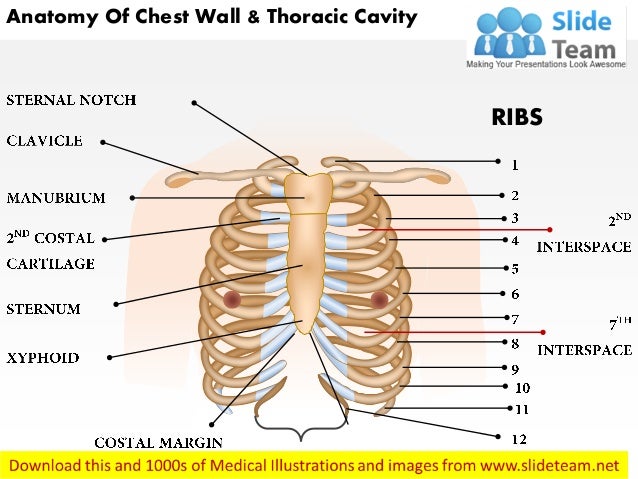
Anatomy of chest wall and thoracic cavity medical images ... from image.slidesharecdn.com Twelve pairs of ribs extend laterally and anteriorly from the thoracic vertebrae to meet at or near the sternum. ■ describe the anatomical relationships of various organs in the chest. The ribs are the bony framework of the thoracic cavity. This type of ct scan uses a lower radiation level than a conventional. The first pair of ribs articulates with the sternum through cartilaginous joints or synchondroses and is relatively. Attach directly to sternum.false ribs: They also have a role in ventilation; Basic rib anatomy consists of a head, neck, tubercle.
The anatomical structure of the 24 ribs in the human body is complex because of the irregular shape and different lengths of each rib.
Spiral ct of thoracic inlet. This type of ct scan uses a lower radiation level than a conventional. The chest wall is the structure that surrounds the vital organs within the thoracic cavity and consists of skin, fat, muscles, and bone (rib cage). The second most common chest wall abnormalities that we see on a cxr are metastases in vertebral bodies and ribs. Moving during chest expansion to enable lung inflation. Don't just draw a generic rib cage shape in there. ■ describe the anatomical relationships of various organs in the chest. The ribs are the bony framework of the thoracic cavity. The rib cage also anchors the bones of the head, neck, shoulders, and arms to the trunk of the body. Twelve pairs of ribs extend laterally and anteriorly from the thoracic vertebrae to meet at or near the sternum. The epidermis is the outermost layer that provides a protective, waterproof seal over the body. Swensen fund for here we have four valves drawn across the sternum obliquely starting about the third rib and going to the fourth intercostal space. Attach directly to sternum.false ribs:
The ribs/costal cartilages have various attachments to the sternum. Twelve pairs of ribs extend laterally and anteriorly from the thoracic vertebrae to meet at or near the sternum. Radiology basics of chest ct anatomy with annotated coronal images and scrollable axial images to help medical students and junior doctors learning anatomy. Increases volume of the chest. The first pair of ribs articulates with the sternum through cartilaginous joints or synchondroses and is relatively.
Types of Chest Trauma and Injuries from www.verywellhealth.com Learn more on this topic. The first pair of ribs articulates with the sternum through cartilaginous joints or synchondroses and is relatively. Anatomy and physiology chest, ribs and respiratory system. Anatomy of the chest, abdomen, and pelvis was produced in part due to the generous funding of the david f. The epidermis is the outermost layer that provides a protective, waterproof seal over the body. Basic rib anatomy consists of a head, neck, tubercle. To determine if patient had good inspiration, what must be seen? True ribs, false ribs, and floating ribs.
Powerful muscles that move the head and arms attach to these bones as well.
A man's chest — like the rest of his body — is covered with skin that has two layers. Radiology basics of chest ct anatomy with annotated coronal images and scrollable axial images to help medical students and junior doctors learning anatomy. Basic rib anatomy consists of a head, neck, tubercle. Insert contains images of a typical rib and the first rib. The ribs/costal cartilages have various attachments to the sternum. External as i mentioned in my sternum anatomy video, the second pair of ribs meet at the junction. They also have a role in ventilation; Identify the following structures on the lateral chest radiograph: The rib cage also anchors the bones of the head, neck, shoulders, and arms to the trunk of the body. Twelve pairs of ribs extend laterally and anteriorly from the thoracic vertebrae to meet at or near the sternum. The first seven ribs attach to the sternum directly and are called true ribs. ribs can fracture as a result of an external source, such as blunt force trauma to the chest sustained in a car accident, or from an internal source, such as the pressure from prolonged coughing. The second most common chest wall abnormalities that we see on a cxr are metastases in vertebral bodies and ribs. Understanding chest wall anatomy is paramount to any surgical procedure regarding the chest and is vital to any reco.
Twelve pairs of ribs extend laterally and anteriorly from the thoracic vertebrae to meet at or near the sternum anatomy of ribs. Construct a robo skelly rib cage and the pelvis using the bucket method.
You have just read the article entitled Anatomy Of Ribs And Chest - Bones in the body Skeletal system 206 bones in the body ... - The second most common chest wall abnormalities that we see on a cxr are metastases in vertebral bodies and ribs.. You can also bookmark this page with the URL : https://suwadihaa.blogspot.com/2021/03/anatomy-of-ribs-and-chest-bones-in-body.html
Share Awesome
Belum ada Komentar untuk "Anatomy Of Ribs And Chest - Bones in the body Skeletal system 206 bones in the body ... - The second most common chest wall abnormalities that we see on a cxr are metastases in vertebral bodies and ribs."

:max_bytes(150000):strip_icc()/GettyImages-186449742-56a05fee3df78cafdaa14dcf.jpg)
Belum ada Komentar untuk "Anatomy Of Ribs And Chest - Bones in the body Skeletal system 206 bones in the body ... - The second most common chest wall abnormalities that we see on a cxr are metastases in vertebral bodies and ribs."
Posting Komentar